Advanced Bionics HiRes™ Bionic Ear System
AB's HiResolution Cochlear Implant Technology
Featuring Two AB Cochlear Implants:
Capturing and delivering details of sound
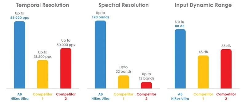
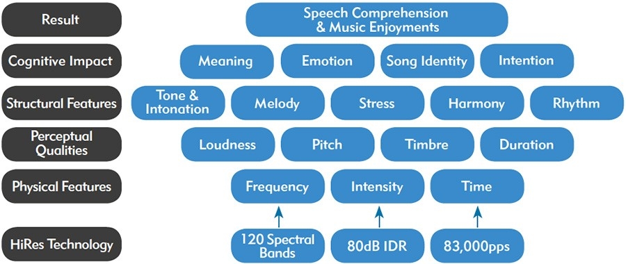
HiResolution implantable technology is the foundation for optimal hearing. The quality of the sound delivered by a cochlear implant system is a direct result of how well the system captures and delivers the details of sound. The HiRes family of cochlear implant was designed to deliver all of the loudness, pitch, and timing information that is essential for natural sound perception and appreciation of music: it automatically encodes the widest range of intensities (up to 80 decibels), it is capable of delivering frequency information to 120 cochlear places using a patented delivery method called current steering, and it provides up to 83,000 pulses per second1.

Current Steering: Hear the Most Subtle Pitch Changes
The number and placement of the actual electrode contacts should not dictate the pitch differences a recipient can detect. Under software control, the 16 independent current sources of the AB implant can steer stimulation to 120 separate locations along the cochlea, thereby increasing the amount of frequency information that can be delivered2. Recipients may take advantage of this enhanced spectral information to hear more pitches, which can improve speech understanding in noise, music appreciation, and tonal language perception.3,4,5 In fact, using psychoacoustic tests, some AB implant recipients can hear as many as 451 distinct pitches with current steering.6
Spiral Ganglion Cells in the Cochlea

Conventional stimulation
Single electrode contact stimulated and
focused on adjacent SG cells
Spiral Ganglion Cells in the Cochlea

Current steering
Two electrode contacts simultaneously stimulated and focused on SG cells between the actual contact locations
Current Steering allows to stimulate the Spiral Ganglion cell population between electrode contacts.
Access to Speech and Music
AB recipients can use fine spectral and temporal information to hear sound accurately, enabling them to better understand tonal information in speech and to enjoy music.6,7,8 An adult will have the best opportunity to reconnect with the hearing world; a child can have access to the best speech and language development possible.9,10,11
Bidirectional Communication Link Between Implant and Sound Processor
All Advanced Bionics recipients or their caregivers can be confident that the implant is functioning properly and that they can benefit from all features of our technology thanks to the proprietary Bidirectional Inductive Communication Link that relays information about the implant’s functional status in real time back to the sound processor. The implant together with the sound processor build a closed loop that ensure proper functioning of the system.
It’s What's Inside That Counts
Internal Components of the AB Cochlear Implant System –
Key Features Explained
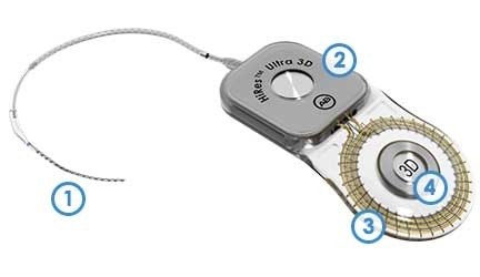
HiRes™ Ultra 3D Cochlear Implant
HiRes™ Ultra 3D Cochlear Implant |
1 Electrode Array
Designed for a gentle cochlear insertion11,12,13 the electrodes deliver 120 spectral bands of sound to help you understand speech and enjoy music.14
2 Electronics Package
Advanced technology designed to support current and future generations of sound processors and features.
3 Communication Link
This sophisticated communication link receives digital representations of sound from the external sound processor and sends information about the status of the implant system and your hearing nerve back to the processor.
4 Innovative Multi-magnet Assembly
The multi-magnet assembly in the HiRes Ultra 3D implant allow users to safely undergo high resolution imaging, such as 3.0 Tesla MRI without removing the multi-magnet assembly. Other implants have differing conditions for conducting safe MRI procedures.
Electrodes Designed for Choice: Without Compromise
The HiRes Ultra implant offers two electrode designs, the straight HiFocus™ SlimJ electrode and the precurved HiFocus™ Mid-Scala electrode, to offer the surgeon a choice based on their practice preferences and the recipient’s anatomy. Both electrodes share the HiFocus design elements.
HiFocus electrode contacts are encased in a slim flexible tapered silicone carrier to minimize insertion forces and damage to cochlear structures during surgery.16,17,18 HiFocus electrodes are designed with balanced stiffness, which allows for easy insertion within the scala tympani while making it less prone to bend upwards towards the basilar membrane and translocate. By minimizing cochlear disruption, HiFocus electrodes offer an increased opportunity for better hearing outcomes.19,20
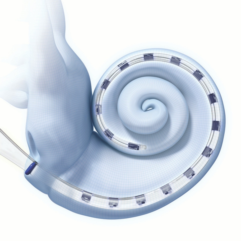
HiFocus SlimJ electrode
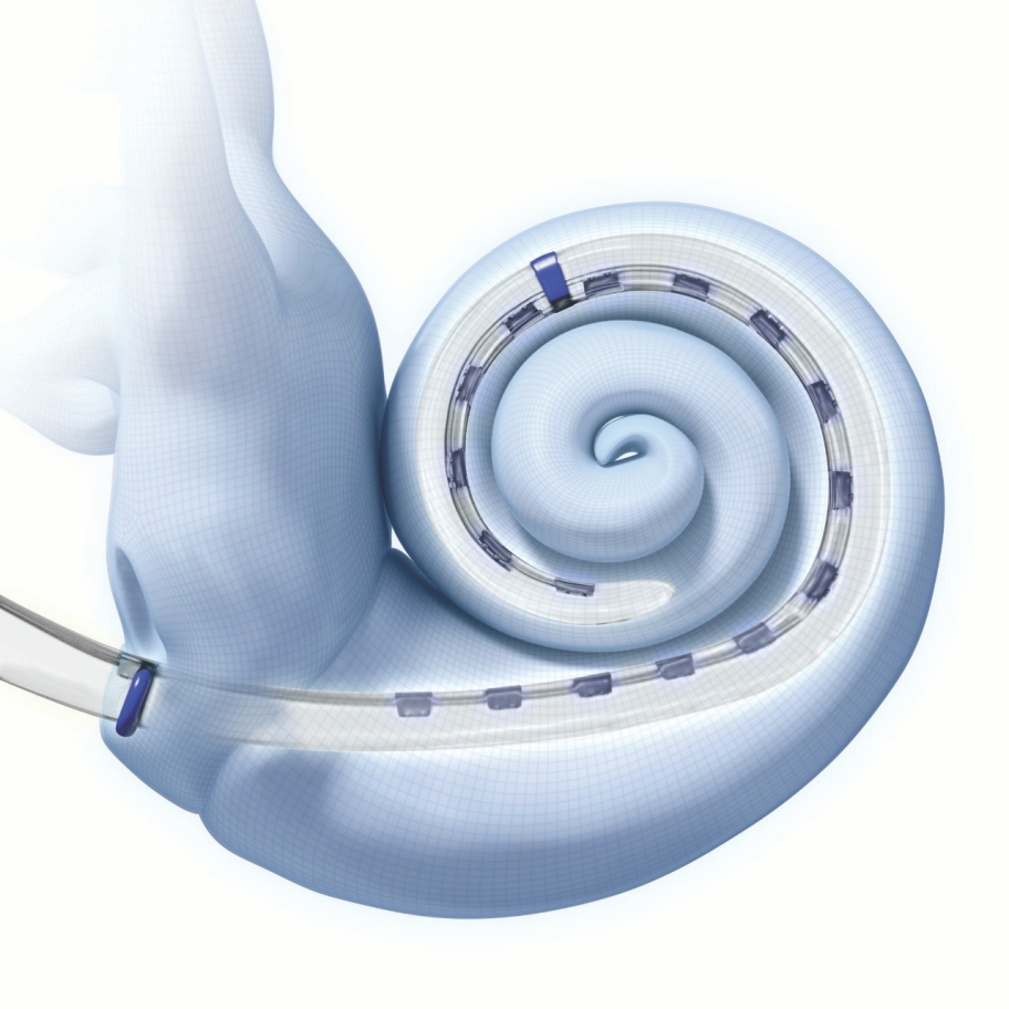
HiFocus Mid-Scala electrode
The HiFocus SlimJ or HiFocus Mid-Scala electrode provide the surgeon with maximum surgical flexibility based upon surgical preference while maintaining patient performance.17,18,19
HiFocus SlimJ
The HiFocus™ SlimJ electrode is the latest approved electrode technology, designed for ease of handling and insertion. It is offered as a straight electrode with a gentle curvature, designed to be easily and smoothly inserted by freehand technique or with forceps. The main benefit of the gentle curvature next to easy insertion is to ensure electrode movement in the apical direction.

1 Balanced stiffness and tapered array profile
Protects cochlear structures.
2 Optimal lateral wing
Allows for ease of handling and insertion of the electrode.
3 Gentle electrode curvature
Ensures electrode movement in the apical direction.
4 Tapered tip feature
For easy and smooth round window insertion.
Confidence of Insertion
Key to the design are the elements that allow a surgeon to easily handle the electrode in the surgical space and insert with minimal trauma to the delicate cochlea structures.17 The HiFocus SlimJ electrode has been designed to have balanced stiffness and flexibility to offer smooth insertion and protect cochlear structures. The wing feature allows for the best possible visualization of the cochlea, and precise control of the angle and speed of insertion. It provides an easy area for a surgeon to hold and control the electrode, even into the facial recess.
The HiFocus SlimJ electrode can be introduced into the cochlea by a surgeon’s preferred approach — by using round window, extended round window, or small cochleostomy, requiring only a 0.8mm opening. The tip feature is intended to ease the insertion through the round window.
“Brilliant electrode design, easy to insert without any resistance maintaining the right orientation, resistant to rotation, and fills the round window beautifully. HiFocus SlimJ is an ideal array for hearing preservation”
- Mr Sherif Khalil, MD, Cochlear Implant Surgeon, UK
Full Spectral Coverage
A marker provides visual indication of insertion depth — the 23mm indicator represents approximately 420° in a standard cochlea, covering the main spiral ganglion population21 to provide optimal spectral coverage.
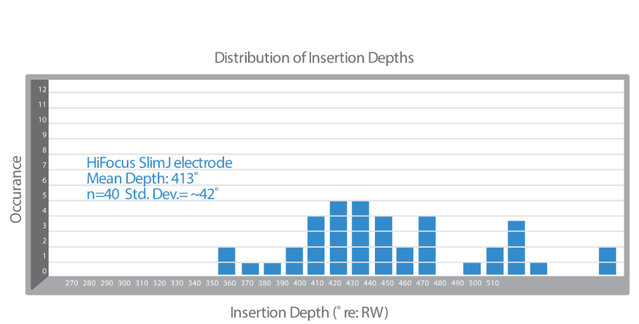
Graph showing angular insertion depths of HiFocus SlimJ across 40 samples17
Cochlear Structure Preservation
Cochlear structure preservation allows for the best possible hearing outcomes in recipients. Studies have shown that recipients may perform better when cochlear structures are undamaged by the electrode insertion.16,19,20,21 The HiFocus SlimJ electrode can be inserted and reinserted up to three times.
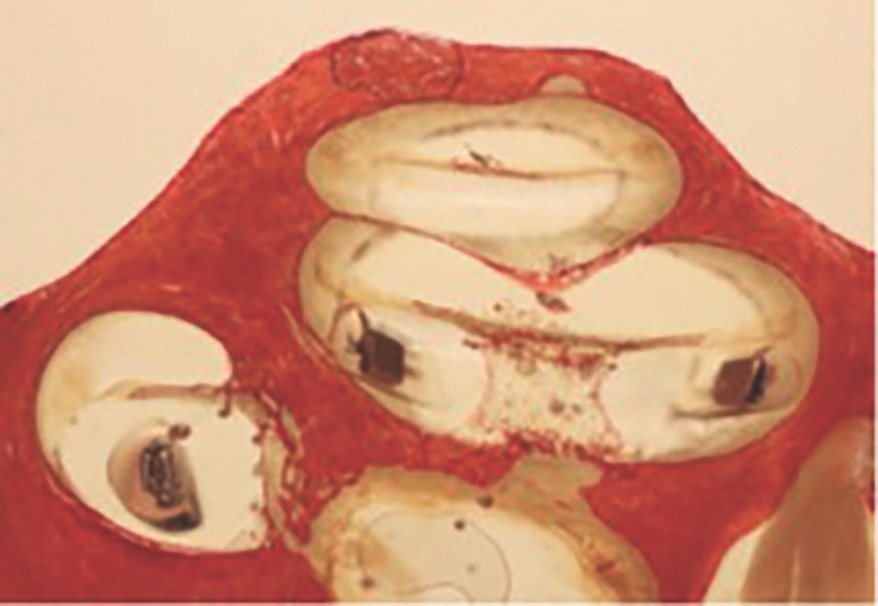
Histology showing HiFocus SlimJ electrode ideally positioned in the Scala Tympani (Eshraghi Scale ‘0’)17
“Based on our multi-center studies in association with investigators at UCSF over the past 18 years, and a review of published reports, the results with the Hifocus SlimJ electrode are remarkable. The HiFocus SlimJ preserves cochlear structures better than any other lateral wall electrode tested to date.”
- Steve Rebscher, Specialist, Department of Otolaryngology, School of Medicine, University of California, San Francisco
HiFocus Mid-Scala
The HiFocus™ Mid-Scala electrode is the smallest styleted precurved electrode designed for consistent positioning in the scala tympani to protect the delicate cochlear structures.
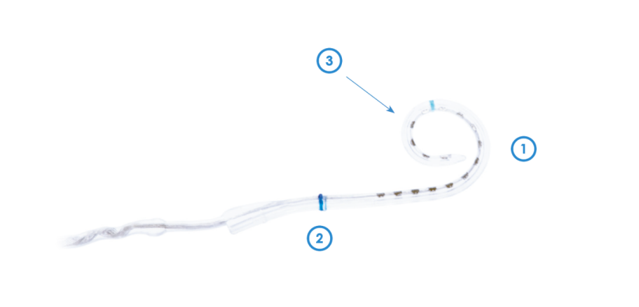
1 One hand insertion
Only pre-curved electrode in the market designed for an easy controlled one hand insertion.
2 Placed mid-scala in the scala tympani
Only electrode in the market designed to be placed mid-scala in the scala tympani.
3 Tapered Tip feature
For Round Window insertion, with Straight Tip region to avoid tip fold over.
Consistency of Placement
Key to the design are the precurved shape, allowing the HiFocus Mid-Scala electrode to be inserted consistently with minimal cochlea trauma,18 a straight tip region to avoid tip fold overs, and if desired, the electrode can be loaded on a dedicated insertion tool to support a controlled insertion.
The HiFocus Mid-Scala electrode can be introduced into the cochlea by a surgeon’s preferred approach — freehand or by use of the insertion tool. It can be inserted using the round window, extended round window, or small cochleostomy approach, requiring only a 0.8mm opening. The tip feature is intended to ease the insertion through the round window
The distal blue marker can be used to ensure the electrode is properly positioned prior to the off stylet technique, thus avoiding tip fold over issues. The proximal blue marker provides a visual indication of a ‘full’ insertion depth — representing approximately 420° angular insertion in a standard cochlea, covering the main spiral ganglion population21 for optimal spectral coverage.
Full Spectral Coverage
The length and curvature of the HiFocus Mid-Scala allows for proven consistency of full spectral coverage with 422° insertion depth signifying coverage of main Spiral Ganglion cell population21 with a tight standard deviation of 20.7°.

Graph showing angular insertion depths of HiFocus Mid-Scala across 35 samples15
Cochlear Structure Preservation
The shape of the HiFocus Mid-Scala places the electrode within the scala tympani, close to the spiral ganglion cells for maximum performance.16,21 The electrode dimensions easily fit within the scala tympani which has been shown to protect the delicate structures of the cochlea18 whilst avoiding damage to the modiolus, osseous spiral lamina and the basilar membrane.18,22,23 HiFocus Mid-Scala, being located central to perimodiolar, has an ideal basal placement for high frequencies.22 The HiFocus Mid-Scala electrode can be inserted and reinserted up to three times.
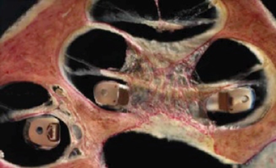
Histology showing HiFocus Mid-Scala electrode ideally positioned in the middle of the scala tympani.
Electrode Options:
References
Ruckenstein, Michael (2012) Cochlear Implants and Other Implantable Hearing Devices
Koch D. B., Downing M., Osberger M. J., and Litvak L. (2007). “ Using current steering to increase spectral resolution in CII and HiRes 90K users,” Ear Hear. 28(2)
Chang YT, Yang HM, Lin YH, Liu SH, Wu JL. Tone discrimination and speech perception benefi t in Mandarin speaking children fi t with HiRes fi delity 120 sound processing. Otol Neurotol. 2009 Sep;30(6):750-7. doi: 10.1097/MAO.0b013e3181b286b2.
Adams D, Ajimsha KM, Barberá MT, Gazibegovic D, Gisbert J, Gómez J, Raveh E, Rocca C, Romanet P, Seebens Y, Zarowski A., Multicentre evaluation of music perception in adult users of Advanced Bionics cochlear implants Cochlear Implants Int. 2014 Jan;15(1):20-6. doi: 10.1179/1754762813Y.0000000032. Epub 2013 Nov 25.
Firszt JB, Holden LK, Reeder RM, Skinner MW.,Speech recognition in cochlear implant recipients: comparison of standard HiRes and HiRes 120 sound processing. Otol Neurotol. 2009 Feb;30(2):146-52. doi: 10.1097/ MAO.0b013e3181924ff8.
Firszt JB, Koch DB, Downing M, Litvak L. (2007) Current steering creates additional pitch percepts in adult cochlear implant recipients. Otology and Neurotology 28:629 -636.
Koch DB, Osberger MJ, Segel P, Kessler D. (2004) HiResolution and conventional sound processing in the HiResolution Bionic Ear: using appropriate outcome measures to assess speech recognition ability. Audiology and Neuro-otology 9:214 -223.
Spahr AJ, Dorman M, Loiselle L. (2007) Performance of patients using different cochlear implant systems: effects of input dynamic range. Ear and Hearing 28(2):26 --275.
Levitin, Daniel (2007) - This is your brain on music, the science of a human obsession
Moira Yip (2002). Tone. (Cambridge Textbooks in Linguistics), Cambridge: Cambridge University Press.
D. Hirst, A. Di Cristo (1998). A survey of intonation systems. In: D. Hirst, A. Di Cristo (Eds.). Intonation Systems, a Survey of Twenty Languages. Cambridge University Press Cambridge (1998)
EN 45502-2-3:2010. Active Implantable Medical Devices. Particular Requirements for Cochlear and Auditory Brainstem Implant Systems.
Internal testing per EN 45502-2-3:2010. Data on fi le.
Internal testing. Data on file.
2017 Cochlear Implant Reliability Report, PN 027-N025-02
Rebscher SJ, Hetherington A, Bonham B, Wardrop P, Whinney D, Leake PA. Considerations for design of future cochlear implant electrode arrays: Electrode array stiffness, size, and depth of insertion. JRRD. 2008 45(5):731-748.
Rivas A, Isaacson B, Kim A, Driscoll C, Cullen R, Rebscher S, (2017) New Lateral Wall Electrode, Evaluation of Surgical Handling, Radiological Placement, and Histological Appraisal of Insertion Trauma , San Francisco, July 26 -29, 2017.
Hassepass F, Bulla S, Maier W, Laszig R, Arndt S, Beck R, Traser L, Aschendorff A; The New Mid-Scala Electrode Array: A Radiologic And Histologic Study In Human Temporal Bones. Otology & Neurotology 2014; 35(8):1415-20
van der Jagt MA1, Briaire JJ, Verbist BM, Frijns JH., Comparison of the HiFocus Mid-Scala and HiFocus 1J Electrode Array: Angular Insertion Depths and Speech Perception Outcomes., Audiol Neurootol. 2016;21(5):316-325. doi: 10.1159/000448581. Epub 2016 Nov 21.
Finley CC, Holden TA, Holden LK, Whiting BR, Chole RA, Neely GJ, Hullar TE, Skinner MW. Role of electrode placement as a contributor to variability in cochlear implant outcomes. Otol Neurotol. 2008 Oct;29(7):920-8.
Avci Ersin, Nauwelaers Tim, Lenarz Thomas, Hamacher Volkmar, Kral Andrej, Variations in microanatomy of the human cochlea, The Journal of Comparative Neurology 2014 Oct 1; 522(14): 3245 -3261.
Gazibegovic D, Bero EM. (2016) Multicenter surgical experience evaluation on the mid-scala electrode and insertion tools. European Archives of Oto- Rhino-Laryngology (Aug 11, epub ahead of print).
Maja Svrakic, J. Thomas Roland Jr, Sean O. McMenomey, and Mario A. Svirsky. Initial Operative Experience and Short-term Hearing Preservation Results With a Mid-scala Cochlear Implant Electrode Array, Otol Neurotol. 2016 Dec;37(10):1549-1554.

